After soaking off our gel polish or rebalancing our enhancements it’s important to inspect our clients nail plates for any signs of abnormalities.
We don’t want to see …
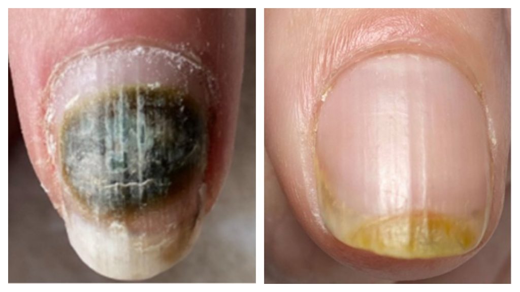
- Yellowing of the nail plate
Esto podría ser onicólisis o daños en la lámina ungueal interna
- Efectos verdosos sobre o bajo la lámina ungueal
Esto podría ser el comienzo de una infección bacteriana
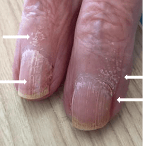
- Zonas blancas calcáreas en la superficie de la uña
This could be white superficial onychomycosis
Los tres nos dan una contraindicación para seguir atendiendo al cliente y los 3 necesitan ser atendidos de forma completamente diferente. (Consulte nuestra Afecciones de las uñas sección)
Pero, ¿cuántos de nosotros miramos detrás de the natural nail at the free edge? Be honest with yourself here – I know my nail tech does but we were trained in the same place so that’s logical.
Quiero enseñarte algo: Onicólisis

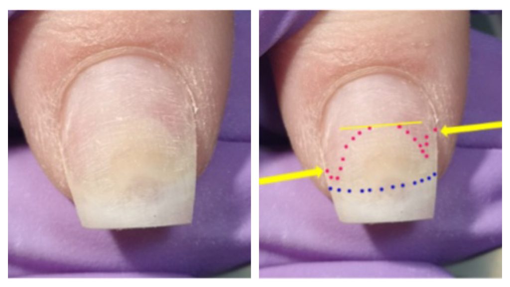
This is what one of my colleges saw when she removed her artificial nail product – I forgive her the missing lateral side walls at the time she didn’t realise they were one of the 4 guardian seals of the nail unit.
The yellow line shows where the nail plate is attached to the nail bed like it should be.
The yellow arrows show where the onycholysis ends but also the amount of detached nail plate in the lateral nail fold.
El rosa señala el extremo proximal de la onicólisis.
Los puntos azules donde banda onicodérmica and the hyponychium should be.
Have a look at this picture of the nail unit from under the free edge. The yellow arrows and line show the detachment of the hyponychium and onychodermal band.
Now imagine if this college was just doing an infill maybe from a dark blue or black hard gel or builder gel polish – no way she would see just how bad the onycholysis is a menos que mirara detrás del borde libre. La onicólisis consume casi 2/3rd of the nail! That’s enough space for a family of bacteria and other mean pathogens to have a vacation in for 6 months!

¿Qué ocurre cuando nos perdemos eso?
¡Esto pasa! Pseudomonas Aeruginosa bacterial infection.

Aquí es donde no quieres estar, este cliente de un salón aquí en Holanda ha estado trabajando durante 6 meses siguiendo el protocolo para reducir esta infección bacteriana, quitar la placa de la uña no era una opción y no lo haríamos a menos que un médico o enfermera especialista nos lo pidiera y entonces sólo bajo condiciones controladas.
Lastly – Onicomicosis superficial blanca

White Superficial Onychomycosis is a surface fungal infection that can affect not only the nails but also the skin – Here in the Netherlands we have a protocol to deal with this infection – please note to nunca file it of the nail plate as it can spread easily, become airborne and possibly enter your lungs.
But – si detectamos antes estos problemas – if we look behind the free edge during rebalancing, if we pay attention during removal we can see and deal with problems before they turn into nightmares.
Sometimes ‘stuff’ just happens, it’s not always something you did or didn’t do, maybe the artificial nail product just cracked and water seeped in and Pseudomonas Aeruginosa (it’s not called an opportunistic pathogen for nothing) had a field day and took hold, maybe the product cracked and she is vulnerable to WSO and it took hold (that’s really possible) and maybe your client has suddenly showed signs of an allergic reaction to your product (keeping away from the skin will help here) but when you don’t know what to look for and you are worried for your client its almost understandable that you just try to cover it up, but please don’t.
Get educated and fix the problem, accidents happen, that’s not the problem, the problem is ignoring them or not having the right information to snap them in the bud.
Don’t only get educated but keep renewing your education, times change, knowledge is expanded all of the time, medical science moves at amazing speed – it’s up to us to keep up!

