Constantemente nos preguntan sobre las diversas características protectoras del lecho ungueal, en particular los roles del hiponiquio y la banda onicodermal. Muchos se preguntan por qué existen dos tipos de sellos y en qué se diferencian del cuerno de la suela.
Esta confusión suele deberse a la falta de claridad de las ilustraciones y explicaciones.
En este artículo pretendemos aclarar estos conceptos explorando las intrincadas estructuras y funciones de estas capas protectoras. Comprender las hiponiquioEl estudio de los factores que contribuyen a la salud y la protección del lecho ungueal, la banda onicodérmica y el cuerno del pie, a menudo incomprendido, proporcionará una visión más clara de su contribución a la salud y la protección general del lecho ungueal.

Aclarar el papel del epitelio del lecho ungueal
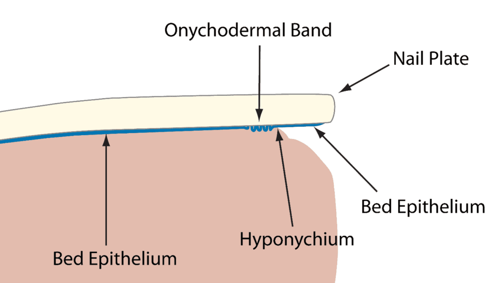
Sé que esto es confuso porque muchas de las ilustraciones actuales no son claras y lo que se necesita es claridad. Intercalado entre la placa ungueal y el lecho ungueal hay una capa de tejido importante, pero muy delgada, llamada epitelio del lecho ungueal.
Muchos no lo entienden, pero esta fina capa de tejido ayuda a guiar la lámina ungueal a medida que crece. Curiosamente, el epitelio del lecho ungueal se adhiere solo a la parte inferior de la lámina ungueal y no al lecho ungueal en sí. Esto permite que la lámina se deslice suavemente sobre el lecho ungueal permitiendo que eventualmente alcance el borde libre y más allá.
A medida que la placa ungueal se mueve, el epitelio del lecho ungueal continúa adhiriéndose firmemente a la parte inferior de la placa ungueal, incluso cuando esta se mueve más allá del dedo y se puede ver en la parte inferior de la uña, todavía firmemente adherida. Este tejido generalmente se elimina durante una manicura al limpiar debajo del borde libre. El tejido firmemente adherido en la parte inferior de la placa ungueal sigue siendo el “epitelio del lecho ungueal”, pero algunos sienten la necesidad de darle un nuevo nombre y lo llaman “solehorn”.
Reevaluando el término “Solehorn”: Por qué la denominación correcta es importante
No estoy de acuerdo con que se use este nombre anticuado. ¿Por qué?
Este término existía antes de que se comprendiera correctamente el verdadero origen de este tejido. El cuerno de la suela no surge de la nada ni procede del hiponiquio, así que ¿por qué habría que darle un nombre diferente? Sigue siendo tejido epitelial del lecho y debe llamarse por su nombre.
Comprender la banda onicodérmica: Un efecto visual del epitelio del lecho
La banda onicodermal NO es otro tipo de tejido a pesar de que forma una región distinta. La banda onicodermal también es creada por el epitelio del lecho ungueal. ¿Cómo? A medida que el epitelio del lecho se mueve con la lámina ungueal, debe de alguna manera pasar por el hiponiquio, que es el sello debajo del borde libre que previene que patógenos infecten el lecho ungueal. Es un paso difícil al pasar por este sello, pero el epitelio del lecho logra pasar. Al hacerlo, se amontona y no fluye suavemente más allá del hiponiquio. Este “amontonamiento” causa un efecto de ondulación que crea una barrera que previene la penetración de sustancias que se filtren más allá del hiponiquio, por lo que esto ayuda a proteger el lecho ungueal.
En la zona que precede al hiponiquio es donde ocurre esta acumulación del lecho epitelial. Este tejido arrugado y abultado produce una zona grisácea llamada banda onicodérmica. Por lo tanto, este es solo un efecto visual causado por la acumulación del lecho epitelial a medida que crece más allá del hiponiquio. No se puede ver la banda onicodérmica mirando debajo del borde libre, no está ahí. Se encuentra debajo de la lámina ungueal y sobre el lecho ungueal y solo se puede ver al mirar a través de la lámina ungueal y observar la banda grisácea que ocurre justo antes del hiponiquio.