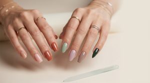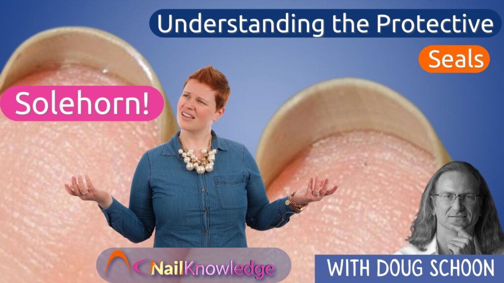
We are constantly asked about the various protective features of the nail bed, particularly the roles of the hyponychium and the onychodermal band. Many wonder why there are two types of seals and how they differ from the solehorn.
This confusion often arises from unclear illustrations and explanations.
In this article, we aim to clarify these concepts by exploring the intricate structures and functions of these protective layers. Understanding the hyponychium, onychodermal band, and the often misunderstood solehorn will provide a clearer picture of how they contribute to the overall health and protection of the nail bed.

Clarifying the Role of Nail Bed Epithelium
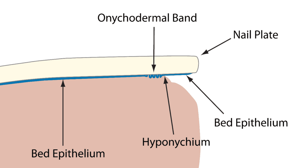
I know this is confusing because many of the current illustrations are unclear and clarity is what is needed. Sandwiched between the nail plate and nail bed is an important, but very thin layer of tissue called the nail bed epithelium.
Many don’t understand, but this paper-thin layer of tissue helps to guide the nail plate as it grows. Interestingly, the bed epithelium adheres only to the underside of the nail plate and not to the nail bed itself. This allows the plate to glide smoothly across the nail bed allowing it to eventually reach the free edge and beyond.
As the nail plate moves, the bed epithelium continues to tightly adhere to the underside of the nail plate, even as it moves past the finger and can be seen on the underside of the nail, still firmly attached. This tissue is usually removed during a manicure when cleaning up under the free edge. The tightly adhered tissue on the underside of the nail plate is still the “bed epithelium”, but some feel a need to give it a new name and they call it “solehorn”.
Reevaluating the Term “Solehorn”: Why Proper Naming Matters
I don’t agree that this old-fashioned name should be used. Why?
This term existed before the true source of this tissue was properly understood. The solehorn does not simply emerge out of nowhere nor does it come from the hyponychium, so why should it be given a different name? This is still bed epithelium tissue and should be called by its proper name.
Understanding the Onychodermal Band: A Visual Effect of the Bed Epithelium
The onychodermal band is NOT another kind of tissue even though it does form a distinct region. The onychodermal band is also created by the bed epithelium. How? As the bed epithelium moves with the nail plate, it must somehow squeeze past the hyponychium, which is the seal under the free edge that prevents pathogens from infecting the nail bed. It’s a tight squeeze getting past this seal, but the bed epithelium manages to squeeze past. In doing so, it becomes bunched up and doesn’t flow smoothly past the hyponychium. This “bunching” causes a rippling effect which creates a barrier that prevents penetration of substances from seeping past the hyponychium, so this helps to protect the nail bed.
In the zone preceding the hyponychium is where this bunch up of the bed epithelium occurs. This bunched up, rippled tissue produces a greyish zone called the onychodermal band. Therefore, this is only a visual effect caused by the bunching up of the bed epithelium as it grows past the hyponychium. You can’t see the onychodermal band by looking under the free edge, it’s not there. It is located underneath the nail plate and on the nail bed and can only be seen by looking though the nail plate and observing the grayish band that occurs just before the hyponychium.