El mayor (¡y mejor!) del mundo Centro de conocimientos sobre uñas

Comprensión de los sellos protectores del hiponiquio del lecho ungueal, la banda onicodérmica y el cuerno de la suela

Comprender los sellos protectores del lecho ungueal

271 visitas

Constantemente nos preguntan por las diversas características de protección del lecho unguealen particular las funciones del hiponiquio y el banda onicodérmica. Muchos se preguntan por qué hay dos tipos de juntas y en qué se diferencian del claxon.

Esta confusión suele deberse a la falta de claridad de las ilustraciones y explicaciones.

En este artículo pretendemos aclarar estos conceptos explorando las intrincadas estructuras y funciones de estas capas protectoras. Comprender las hiponiquioEl estudio de los factores que contribuyen a la salud y la protección del lecho ungueal, la banda onicodérmica y el cuerno del pie, a menudo incomprendido, proporcionará una visión más clara de su contribución a la salud y la protección general del lecho ungueal.

Aclarar el papel del epitelio del lecho ungueal
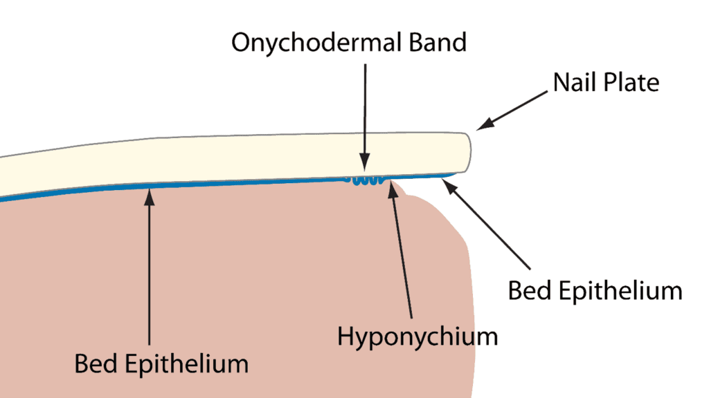
Sellado protector del lecho ungueal

Sé que es confuso porque muchas de las ilustraciones actuales no son claras y lo que hace falta es claridad. Entre el placa para clavos y el lecho ungueal hay una capa de tejido importante, pero muy fina, llamada epitelio del lecho ungueal

Muchos no lo entienden, pero esta fina capa de tejido ayuda a guiar la lámina ungueal a medida que crece. Curiosamente, la epitelio del lecho se adhiere únicamente a la parte inferior de la placa ungueal y no al lecho ungueal propiamente dicho. De este modo, la placa se desliza suavemente por el lecho ungueal y llega a la uña. borde libre y más allá. 

A medida que la placa ungueal se mueve, el lecho epitelio sigue firmemente adherido a la parte inferior de la lámina ungueal, incluso cuando se desplaza más allá del dedo y puede verse en la parte inferior de la uña, todavía firmemente adherido. Este tejido suele eliminarse durante una manicura al limpiar debajo del borde libre. El tejido fuertemente adherido de la parte inferior de la lámina ungueal sigue siendo el "epitelio del lecho", pero algunos sienten la necesidad de darle un nuevo nombre y lo llaman "cuerno de la suela". 

Reevaluación del término "Solehorn": Por qué es importante nombrarlo correctamente

No estoy de acuerdo en que se utilice este nombre anticuado. ¿Por qué?

Este término existía antes de que se comprendiera correctamente el verdadero origen de este tejido. El cuerno de la suela no surge de la nada ni procede del hiponiquio, así que ¿por qué habría que darle un nombre diferente? Sigue siendo tejido epitelial del lecho y debe llamarse por su nombre.
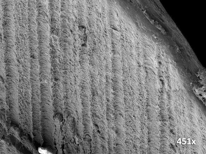
Epitelio de lecho adherido a la cara inferior del borde libre.

Comprender la banda onicodérmica: Un efecto visual del epitelio del lecho

La banda onicodérmica NO es otro tipo de tejido, aunque sí forma una región diferenciada. La banda onicodérmica también es creada por el epitelio del lecho. ¿Cómo? Cuando el epitelio del lecho ungueal se mueve con la lámina ungueal, de alguna manera debe pasar a través del hiponiquio, que es el sello situado bajo el borde libre que impide que la lámina ungueal se abra. patógenos de infectar el lecho ungueal. El epitelio del lecho ungueal se las arregla para pasar. Al hacerlo, se amontona y no fluye suavemente más allá del hiponiquio. Este "amontonamiento" provoca un efecto de ondulación que crea una barrera que impide la penetración de sustancias a través del hiponiquio, lo que ayuda a proteger el lecho ungueal.

En la zona que precede al hiponiquio es donde se produce este agrupamiento del epitelio del lecho. Este tejido agrupado y ondulado produce una zona grisácea denominada banda onicodérmica. Por lo tanto, se trata únicamente de un efecto visual causado por el amontonamiento del epitelio del lecho a medida que crece más allá del hiponiquio. No se puede ver la banda onicodérmica mirando por debajo del borde libre, no está ahí. Se encuentra debajo de la lámina ungueal y en el lecho ungueal y sólo se puede ver mirando a través de la lámina ungueal y observando la banda grisácea que se produce justo antes del hiponiquio.

es_ESSpanish
Cesta de la compra
\n

Thin epithelium tissue on top of the nail bed dermis and under the nail plate, which allows nail plate to glide across, while remaining strongly attached to the underlying nail bed through interlocking ridges and grooves.<\/p>\n<\/div>","cmtt_519a81190b04808191db83dcdfbdfcff":"

Onychodermal band<\/div>
Onychodermal Band\n

A smile line showing through the Nail Plate which is caused by Nail Bed Epithelium squeezing past the Hyponychium.<\/p>\n<\/div>","cmtt_9f16b70f5361b0cd305163ba5f6389c7":"

Bed Epithelium<\/div>
Nail Bed Epithelium\n

This is a thin layer of specialised epithelial tissue that sits on the nail bed and under the nail plate.

At the proximal end it is connected to the nail matrix. At the distal end it forms part of the hyponychium seal.

The onychodermal band is where it squeezes past this seal. It is also involved in the condition: hyperkeratosis which is a reaction to several aggressive factors.<\/p>\n<\/div>","cmtt_ab4313011d592bef769a0612daf67df9":"

Hyponychium<\/div>
The Hyponychium\n

The Hyponychium is the nail seal at the distal edge of the nail bed. A slightly thickened layer of skin, it lies beneath the free edge of the nail plate.<\/p>\n<\/div>","cmtt_1246a1b7fb9bed3e4702bd0168f71114":"

Nail plate<\/div>
Nail Plate\n

Hard layers of modified and keratinised skin cells, bonded together, to form a protective plate on the end of each finger and toe, that prevents damage to the underlying bone and allows for manual dexterity.<\/p>\n<\/div>","cmtt_3862ec5812109e89f2ef491358667ded":"

Free edge<\/div>
\n

The free edge refers to the outermost part of the nail plate that extends beyond the fingertip or toe. It is the portion of the nail that is not attached to the nail bed or the surrounding skin. The free edge is typically trimmed and shaped during nail care routines and is susceptible to snapping or breaking off when subjected to excessive force or trauma.<\/p>\n<\/div>","cmtt_b3f25bf5945a3ed561fe93c3d08ef964":"

Epithelium<\/div>
\n

Epithelium is a type of tissue that forms the outermost layer of the body's organs, as well as the inner lining of cavities and surfaces throughout the body. It serves as a protective barrier and can have various shapes and functions depending on its location, such as absorbing nutrients in the intestines, secreting mucus in the respiratory tract, or providing a protective layer for the skin.<\/p>\n
\n

Usage: \"The epithelium of the skin acts as a protective barrier against external factors, while the epithelium in the small intestine is specialized for nutrient absorption.\"<\/p>\n<\/div>","cmtt_c1a36df0013019a12f8f575de70e853d":"

Nail bed<\/div>
Nail Bed\n

An area of the nail unit under the nail plate consisting of Nail Bed Epithelium and Dermis.<\/p>\n<\/div>","cmtt_e4531ac60c2cb1fbabc9f3c62c5f342c":"

Manicure<\/div>
Nail Technician at Work\n

A manicure is a treatment to help improve the condition and appearance of the hands and fingernails. Usually consists of cuticle removal, shaping of the natural nail's free edge, moisturizing, and color or high shine on the nail.<\/p>\n<\/div>","cmtt_b1c0d6ac2b2d57b70735cc3036131694":"

Pathogen<\/div>
Pathogen\n

A pathogen is the term used to describe an organism (any organism) that can produce disease. <\/p>\n
\n

Viruses, Bacteria\u00a0and Fungi\u00a0are 3 typical pathogens that we can come into contact within the nail salon or podiatry practice.<\/p>\n<\/div>","cmtt_17d90b8db559353662dd528fbaadc71a":"

Seals<\/div>
The Nail Seals of the nail unit\n

The nail plate is sealed all the way around it to protect the nail bed and the nail matrix. The seals are at the proximal nail fold, the side walls (perionychium) and under the free edge of the nail (the hyponychium).<\/p>\n<\/div>"}}; -->