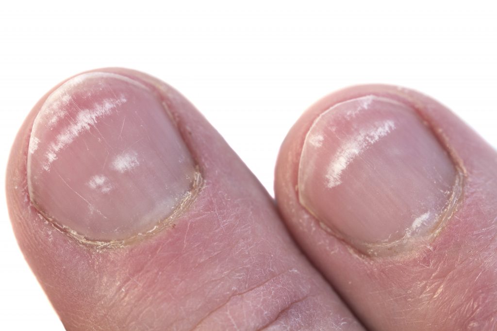
Search for answers you are looking for.
Leukonychia
2.9k Views
What is Leukonychia and What Causes It?
Leukonychia is the clinical term used to describe white discoloration of the nail plate. The word is derived from the Greek leuko (meaning “white”) and onyx (meaning “nail”). In practice, leukonychia may present as white spots, lines, or full whitening of the nail plate and is sometimes referred to informally as “white spots” or “white marks” on the nails.
A disruption in the keratinisation process within the nail matrix, where nail plate cells are formed, results in the condition known as leukonychia. When the process of keratin synthesis is incomplete or disrupted, due to trauma, chemical interference, or systemic health issues, the resulting nail cells may retain their nuclei or form abnormally. These defective cells scatter light differently, producing the white appearance seen in leukonychia.
Depending on which part of the matrix is affected, leukonychia may occur in any area of the nail plate and can involve one, two, or all three keratin layers. This variability explains why leukonychia can appear in different forms and in various locations on the nail surface.
Understanding the underlying cause and classification of leukonychia is crucial in assessing nail health and determining appropriate care or referral.
Classification of Leukonychia
Historically, leukonychia was first classified by Unna in 1896 into:
- Transversal
- Longitudinal
- Punctate
Later, Weber added a fourth category:
- Partialis
To further simplify, leukonychia is now generally divided into the following groups:
- True Leukonychia – due to defects within the nail plate itself
- Apparent Leukonychia – due to issues with the underlying subungual tissue (e.g., nail bed)
- Pseudo Leukonychia – typically caused by superficial fungal infections affecting the surface of the nail
Subtypes of True Leukonychia
True leukonychia can be further categorised into:
- Total – the entire nail plate is white
- Subtotal – most of the nail plate is affected
- Partial – presents as lines, spots, or distal changes originating from localized matrix damage
Types of Leukonychia Seen in Practice
1. Spotted Leukonychia (Punctate/Partial)
Spotted Leukonychia appears as white spots caused by abnormal keratinisation in the proximal matrix. It is common across all age groups.
- The white spots remain when pressure is applied
- Spots grow out with the nail
- Usually harmless and not associated with systemic disease
Do:
- Keep nails short
- Apply oil and lotion
- Reassure the client if the nail plate is intact
Don’t:
- File out the white spots
- Rough up the nail plate

Note:
There is no contraindication for nail products if the nail plate is intact.
2. Striped Leukonychia (Leukonychia Striata)
Stripes may appear longitudinally or transversely. This group includes Muehrcke’s lines and Mees’ lines.
2a. Muehrcke’s Lines (Apparent Leukonychia)
- Bands follow the shape of the lunula
- Caused by chemical disruption to the distal nail matrix
- Do not move with nail growth
- Disappear under digital pressure
- Often seen during chemotherapy or immunotherapy
Do:
- Keep nails short
- Apply oil and lotion
- Refer to a medical professional if needed
Don’t:
- Disturb the nail plate structure

Important:
Contraindication for artificial nails if the immune system is compromised. If artificial enhancements are used, flexible, clear products are advised to reduce pressure on the nail bed.
2b. Mees’ Lines (True Leukonychia)
- Do not disappear with pressure
- Move out with nail growth
- Indicative of underlying systemic issues (e.g., arsenic poisoning, severe illness)
- Require medical evaluation
Do:
- Keep nails short
- Apply oil and lotion
- Refer to a medical professional
Don’t:
- Apply products until medical clearance is obtained

Important:
Contraindicated for artificial nail products until a healthcare provider has cleared the client.
3. Total Leukonychia (Leukonychia Totalis)
- The entire nail plate appears opaque, white, sometimes resembling fine porcelain
- Can be hereditary or caused by abnormal keratinisation in the matrix
- May result from chemo/immunotherapy, arsenic poisoning, or chronic trauma
- The nail may grow more slowly and involve periungual skin changes
Do:
- Keep nails short
- Apply oil and lotion
- Refer to a medical professional if changes in the nail occur
Don’t:
- Disturb the structure of the nail plate

Note:
No contraindication for artificial nail products if the nail plate is structurally intact.
4. Pseudo Leukonychia (White Superficial Onychomycosis)
This condition mimics leukonychia but is caused by superficial fungal infection, commonly by Trichophyton mentagrophytes.
- Appears as opaque, friable, white spots
- Often affects both nails and surrounding skin
- May feel chalky and brittle
- Common in immunocompromised individuals
Do:
- Keep nails clean
- Wash hands with soap and water
- Use anti-fungal spray and quality nail oil
- Refer to a doctor if symptoms persist
Don’t:
- File the white spots
- Cover with artificial nail products
- Ignore the signs

Important:
Do not apply artificial nail products. The nail plate’s porosity may increase the risk of secondary infection.
Rare Forms of Leukonychia
Terry’s Nails
- Form of total leukonychia
- Strongly associated with systemic diseases such as liver failure or diabetes
- The entire nail plate appears white with a narrow pink or brown distal band
Lindsay’s Nails (Half-and-Half Nails)
- Proximal nail appears white
- Distal band occupies 20–60% of nail bed and appears pink, red, or brown
- Linked with renal disease
The Role of Nail Professionals
Leukonychia is sometimes caused by mechanical trauma to the proximal nail fold or nail matrix, resulting in parakeratotic onychocytes — abnormal nail cells that reflect light and appear white.
Best Practices for Nail Professionals:
- Avoid roughing up the nail plate unnecessarily
- Learn to identify different types of leukonychia
- Make informed decisions on product application
- Refer to medical professionals when in doubt
Recognising and understanding leukonychia is critical to client safety, trust, and long-term nail health.
Summary
- Leukonychia can be true, apparent, or pseudo, depending on the origin
- Always investigate the cause before applying nail products
- Some forms are harmless; others may indicate serious health concerns
- Education and careful observation are key to appropriate care
“There is always a reason for any form of leukonychia in or under the nail plate. A well-trained nail professional will recognise and act accordingly.”
References:
Bhoi AK, Gaurav V, Kushwaha S. Leukonychia Totalis: An Underreported Sign of Chronic Arsenicosis. Skin Appendage Disord. 2024 Aug;10(4):325-328. doi: 10.1159/000538450. Epub 2024 Apr 12. PMID: 39108548; PMCID: PMC11299963.
J. C. R. Bowling, S. McIntosh, K. L. Agnew, Transverse leukonychia of the fingernail following proximal nail fold trauma, Clinical and Experimental Dermatology, Volume 29, Issue 1, 1 January 2004, Page 96, https://doi.org/10.1111/j.1365-2230.2004.01432.x
Iorizzo M, Starace M, Pasch MC. Leukonychia: What Can White Nails Tell Us? Am J Clin Dermatol. 2022 Mar;23(2):177-193. doi: 10.1007/s40257-022-00671-6. Epub 2022 Feb 2. PMID: 35112320; PMCID: PMC8809498.
Fernandez-Nieto D, Jimenez-Cauhe J, Ortega-Quijano D, Diaz-Guimaraens B, Dominguez-Santas M, Martinez-Rubio J. Transverse leukonychia (Mees’ lines) nail alterations in a COVID-19 patient. Dermatol Ther. 2020 Nov;33(6):e13863. doi: 10.1111/dth.13863. Epub 2020 Jul 7. PMID: 32779847; PMCID: PMC7323062.
Wagner G, Meyer V, Sachse MM. Leukonychia totalis [Leukonychia totalis]. Hautarzt. 2016 Apr;67(4):308-10. German. doi: 10.1007/s00105-015-3736-4. PMID: 26676417.
Conway J, Lipner SR. Onychomycosis Presenting as Longitudinal Leukonychia. J Cutan Med Surg. 2023 May-Jun;27(3):298. doi: 10.1177/12034754231159598. Epub 2023 Feb 27. PMID: 36851881.
Stevens KR, Leis PF, Peters S, Baer S, Orengo I. Congenital leukonychia. J Am Acad Dermatol. 1998 Sep;39(3):509-12. doi: 10.1016/s0190-9622(98)70341-x. PMID: 9738798.
Dasanu CA, Ichim TE, Alexandrescu DT. Muehrcke’s lines (Leukonychia striata) due to transretinoic acid therapy for acute promyelocytic leukemia. Journal of Oncology Pharmacy Practice. 2013;19(4):377-379. doi:10.1177/1078155212468923
Image gallery


